Combined Micro- and Macro scale X-ray powder diffraction mapping
Combined Micro- and Macro scale X-ray powder diffraction mapping
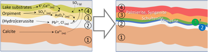
The spontaneous chemical alteration of artists’ pigment materials may be caused by several degradation processes. Some of these are well known while others are still in need of more detailed investigation and documentation. These changes often become apparent as color modifications, either caused by a change in the oxidation state in the original material or the formation of degradation products or salts, via simple or more complex, multistep reactions. Arsenic-based pigments such as orpiment (As2S3) or realgar (α-As4S4) are prone to such alterations and are often described as easily oxidizing upon exposure to light. Macroscopic X-ray powder diffraction (MA-XRPD) imaging on a sub area of a still life painting by the 17th century Dutch painter Martinus Nellius was employed in combination with microscopic (μ-) XRPD imaging of a paint cross section taken in the area imaged by MA-XRPD. In this way, the in situ formation of secondary metal arsenate and sulfate species and their migration through the paint layer stack they originate from could be visualized. In the areas originally painted with orpiment, it could be shown that several secondary minerals such as schultenite (PbHAsO4), mimetite (Pb5(AsO4)3Cl), palmierite (K2Pb(SO4)2) and syngenite (K2Ca(SO4)2∙H2O) have formed. Closer inspection of the cross-sectioned paint layer stack with μ-XRPD illustrates that the arsenate minerals schultenite and mimetite have precipitated at the interface between the orpiment layer and the layer below that is rich in lead white, i.e. close to the depth of formation of the arsenate ions. The sulfate palmierite has mostly precipitated at the surface and upper layers of the painting.

Tandem Probe Analysis Mode for Synchrotron XFM: Doubling Throughput Capacity

Macroscopic x-ray powder diffraction imaging reveals Vermeer's discriminating use of lead white pigments in Girl with a Pearl Earring

X‐ray Diffraction Mapping for Cultural Heritage Science: a Review of Experimental Configurations and Applications - Gonzalez - 2020 - Chemistry – A European Journal - Wiley Online Library

Carbon fibre lattice strain mapping via microfocus synchrotron X-ray diffraction of a reinforced composite - ScienceDirect

Mapping of Structural Changes Induced by X-ray Nanopatterning via Nano-X-ray Diffraction and Corresponding Electrical Effects

Transmission and Reflection Mode Macroscopic X-ray Powder Diffraction Imaging for the Noninvasive Visualization of Paint Degradation in Still Life Paintings by Jan Davidsz. de Heem

Imaging of Biological Materials and Cells by X-ray Scattering and Diffraction
Powder diffraction Nature Reviews Methods Primers
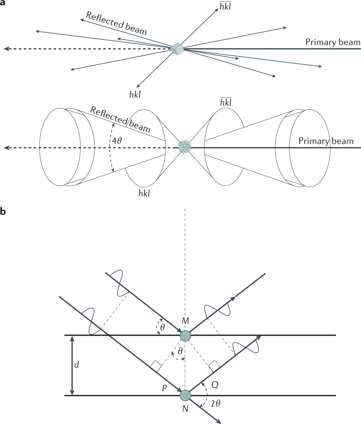
General principle of an XRD mapping experiment. XRD patterns are

X‐ray Diffraction Mapping for Cultural Heritage Science: a Review of Experimental Configurations and Applications - Gonzalez - 2020 - Chemistry – A European Journal - Wiley Online Library
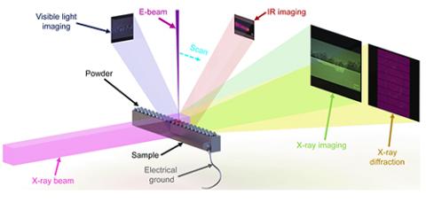
Imaging (XSD-IMG) Advanced Photon Source
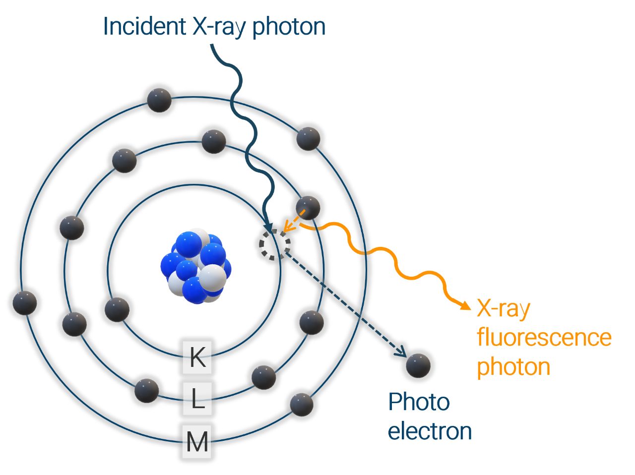
How does XRF work?

PDF] XRDUA: crystalline phase distribution maps by two‐dimensional scanning and tomographic (micro) X‐ray powder diffraction


